Sunspots
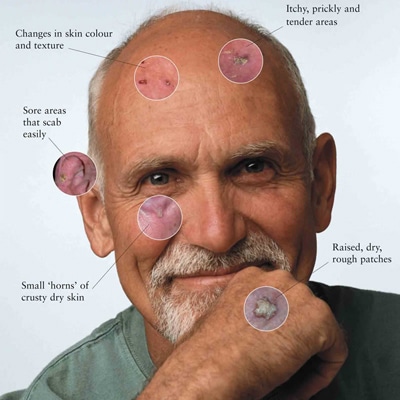
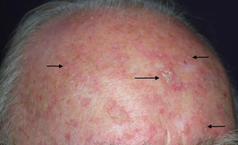
Solar keratosis or sunspots are precancerous skin condition that results from overexposure to the sun. These small rough spots are pink, red or flesh-coloured and often appear on sun-exposed areas of the skin, such as the face, ears, back of the hands and arms.
Solar keratoses may progress to invasive skin cancers, but more importantly serve as a marker for patients who are at a higher risk of developing invasive skin cancers elsewhere on their bodies.
If you noticed these lesions anywhere on your body, it is important that you have your skin checked by a doctor as soon as possible.
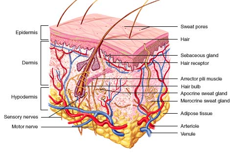
Solar keratoses are pre-cancerous, which means that they have the potential to progress to a type of skin cancer called Squamous Cell Carcinoma (SCC) if they are not treated. SCC is a type of skin cancer that develops due to the uncontrolled growth of abnormal squamous cells. These are the cells that make up the upper layer of your skin (epidermis).
At early stages it usually remains confined to the epidermis but if left untreated, may eventually invade deeper layers of skin. In some people the cancerous cells may spread to distant organs. If this happens, SCC can be fatal. In most cases, invasive SCC results from a solar keratosis lesion.

What is the cause of Solar keratosis?
Solar keratosis results from excessive, long term exposure to UV radiation. This means that people who work outdoors or enjoy outdoor hobbies are at greatest risk of developing solar keratoses.
In general, solar keratoses are most likely to develop in fair skinned people. Older men with baldness are at particularly higher risk of solar keratoses. People who are immunosuppressed or are taking medications that cause them to be immunosuppressed ( such as medications prescribed for organ transplant, rheumatoid arthritis, inflammatory bowel disease ) are also at high risk of developing solar keratoses.

diangnosis
Solar Keratosis is usually diagnosed by its physical appearance on a routine skin check. In some cases, we may need to examine the lesion more closely using a tool known as dermatoscope. Sometimes, we may also need to take a biopsy of the solar keratosis. This involves removing a small sample of the lesion and examining it under a microscope.

Treatment
There are several reasons to treat a solar keratosis, for cosmetic reasons, symptom relief and most importantly, to prevent the lesion from progressing to skin cancer. Some lesions can be left alone and simply keep an eye on them. If the lesions changes in appearance or new lesions appear, we may need to treat them
1.Sugery : Under local anaesthesia, lesion is removed with a scalpel and wound is closed with stitches. The surgical procedure may leave a permanent scar but it is very effective approach to treatment.
2. Curettage: If you have multiple isolated or thick lesions, curettage may be a suitable treatment option for you. During this procedure, abnormal skin is scraped off with a curette (a sharp, spoon like instrument). You will be given a local anaesthesia before the procedure and electrosurgery will be used to stop any bleeding after the lesion has been removed. A scab will be formed after the curettage which will heal over after a few weeks and may leave a small scar.
3. Cryotherapy: Freezing with liquid nitrogen is a common treatment for solar keratosis in Australia. If you have thin and isolated lesions, cryotherapy might be suitable. During this procedure, liquid nitrogen will be sprayed onto your skin lesion. This leads to blistering and shedding of the abnormal skin. Freezing may cause some pain but fortunately a short freeze is usually enough to treat most solar keratoses. This may leave mild scar and hypo pigmentation ( whitening) of treated areas. This will be more noticeable in darker skinned people.
4. Creams: If you have multiple lesions, topical creams or gels might be a suitable treatment option. The location of the lesions will determine how long it take for the treatment to have an effect. Facial lesions respond to treatment more quickly than lesions on arms. Topical agents cause the abnormal skin cells to die but in the process may make your skin sore. Your skin may also weep and blister for a few days after treatment. After topical treatment, there is a chance that solar keratosis may re-occur on the treated area requiring further treatment.
5. PDT If you have multiple thin solar keratosis on face, PDT is a suitable treatment. This form of therapy is associated with better cosmetic results. Before you begin PDT, we will apply a cream to the affected skin. This cream contains a light-activated medicine that will work to destroy the abnormal skin. PDT can be painful for some patients. After the procedure you will notice crusting, redness and blistering of your skin. These are expected side effects and will subside with time. While your skin is healing. It is important to avoid sunlight exposure.


